Advancements in medical imaging have significantly improved the way healthcare professionals monitor skin health and identify subtle changes over time. Among the most valuable innovations in dermatology is photographic mole mapping, a technology-driven approach that allows doctors to document and compare skin lesions with greater precision. Patients seeking a routine skin check sunshine coast are increasingly encountering this modern method during preventive skin assessments, especially when long-term monitoring is recommended.
Photographic mole mapping has transformed skin examinations from simple visual observations into highly detailed tracking systems that help detect evolving skin abnormalities earlier and more accurately. As awareness around preventive care continues to grow, this technology is becoming an essential part of modern skin health management.
Understanding What Photographic Mole Mapping Involves
Photographic mole mapping is a process that captures high-resolution images of a patient’s skin and existing moles for ongoing comparison during future appointments. Instead of relying solely on memory or written descriptions, doctors can evaluate exact visual records over time.
The procedure often includes:
- Full-body photography
- Close-up imaging of individual moles
- Digital storage of skin records
- Comparative analysis during follow-up visits
- Monitoring for subtle visual changes
This system allows clinicians to identify developments that might otherwise go unnoticed during standard examinations.
Why Traditional Skin Monitoring Had Limitations
Before advanced imaging technology became widely available, skin checks relied heavily on manual observation and patient recollection. While experienced doctors could still identify suspicious lesions, tracking gradual changes across months or years was more challenging.
Common Difficulties With Older Monitoring Methods
Human Memory Is Imperfect
Patients may struggle to remember exactly how a mole looked during previous visits.
Subtle Changes Are Easy to Miss
Minor shifts in color, border shape, or size may develop slowly and remain difficult to detect without side-by-side comparisons.
Large Numbers of Moles Increase Complexity
Individuals with many moles often require more detailed tracking than visual observation alone can provide.
Photographic mole mapping helps overcome these limitations by creating a consistent visual reference system.
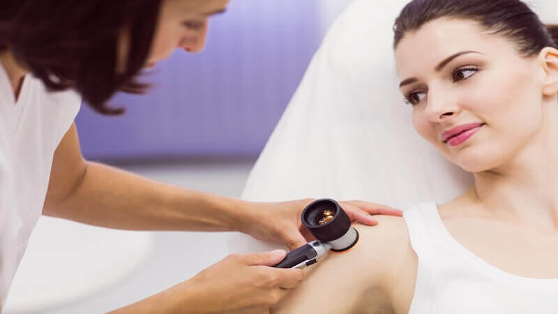
How Doctors Use Mole Mapping During Skin Assessments
During a consultation, doctors review current skin images alongside previous records to look for evolving patterns or abnormalities. This comparative approach provides greater accuracy than isolated observations.
Key Features Doctors Commonly Evaluate
Changes in Shape
Irregular or asymmetrical growth patterns may attract closer attention over time.
Variations in Pigmentation
Differences in color distribution can sometimes indicate abnormal skin activity.
Border Development
Edges that become uneven or blurred may require further investigation.
Growth Progression
Even small increases in lesion size can become more noticeable through image comparisons.
Because photographic records provide objective visual evidence, doctors can make more informed clinical decisions.
The Role of Technology in Early Detection
One of the most important benefits of mole mapping is its contribution to earlier detection. Skin abnormalities often evolve gradually, making small changes difficult to identify during isolated appointments.
With digital imaging, doctors can identify trends that emerge over time rather than relying solely on what is visible in a single visit.
Why Early Detection Matters
Early identification of suspicious lesions may:
- Reduce the need for extensive treatment
- Improve management outcomes
- Allow quicker medical intervention
- Minimize uncertainty during monitoring
- Support more accurate diagnoses
This proactive approach reflects the broader shift toward preventive healthcare across modern medicine.
Why Patients Often Feel More Reassured
Many patients appreciate the clarity and transparency photographic mole mapping provides. Instead of vague descriptions about whether a spot has changed, they can see visual comparisons directly.
This process often helps reduce anxiety because monitoring becomes more measurable and evidence-based.
Patients frequently report greater confidence in:
- Routine skin examinations
- Follow-up recommendations
- Understanding their skin patterns
- Identifying new or evolving lesions
The visual nature of the technology also makes communication between doctors and patients more effective.
Who Benefits Most From Mole Mapping?
While photographic monitoring can benefit many individuals, certain groups often gain particular value from this technology.
People With Numerous Moles
Tracking many skin lesions manually can become difficult without photographic assistance.
Individuals With High Sun Exposure
Outdoor workers and people living in sunny climates may require closer long-term observation.
Patients With Family History Concerns
Those with hereditary skin cancer risk factors often undergo more frequent monitoring.
Individuals With Previous Skin Abnormalities
Patients who have previously experienced suspicious lesions may benefit from ongoing comparative imaging.
Doctors determine whether mole mapping is appropriate based on individual skin characteristics and medical history.
How Mole Mapping Supports Long-Term Skin Awareness
Another advantage of photographic monitoring is the way it encourages patients to become more aware of their own skin health. Seeing documented images over time often motivates individuals to pay closer attention to new or changing spots between appointments.
This increased awareness can improve self-monitoring habits and encourage earlier medical consultation when unusual changes appear.
Everyday Awareness Still Matters
Although advanced imaging is highly valuable, doctors still encourage patients to:
- Perform regular self-checks
- Use sun protection consistently
- Monitor unusual skin changes
- Attend scheduled follow-up visits
- Report symptoms such as itching or bleeding
Technology works best when combined with active patient participation and preventive habits.
The Future of Preventive Skin Monitoring
As digital healthcare technology continues advancing, photographic mole mapping is likely to become even more sophisticated. Artificial intelligence, improved imaging software, and enhanced diagnostic tools are already beginning to support dermatology practices worldwide.
These innovations may eventually help doctors identify suspicious lesions with even greater speed and precision.
At the same time, the core purpose remains unchanged: helping patients monitor their skin more effectively and supporting earlier detection through consistent observation.
A Smarter Approach to Monitoring Skin Over Time
Photographic mole mapping has reshaped the way doctors evaluate skin changes by providing detailed visual records that improve accuracy, consistency, and long-term monitoring. Rather than relying solely on memory or isolated observations, clinicians can now track skin developments through precise image comparisons over months and years.
For patients, this technology offers reassurance, greater awareness, and a more proactive approach to preventive care. As skin health awareness continues growing, photographic monitoring is becoming an increasingly valuable tool in helping people stay informed about changes that might otherwise go unnoticed.
By combining advanced imaging with regular professional assessments and everyday sun protection habits, modern skin care has entered a far more precise and preventive era.